First procedure – cartilage biopsy harvest
0
1. Preparation
- The patient is prepared for arthroscopy, a short and minimally invasive procedure.
- The surgical site is cleaned, and local or general anaesthesia is administered.
2. Procedure (30 minutes)
- Small incisions, usually two or three, are made in the front part of the knee joint.
- An arthroscope, a small camera, is inserted to visualise the inside of the joint.
- The cartilage defect is located, cleaned and measured.
- Healthy cartilage tissue from a non-weight-bearing area of the damaged knee is identified.
- A small cartilage biopsy sample is harvested using special instruments and placed in a specialised transport solution.
3. End of procedure and recovery
- The incisions are closed using stitches or staples.
- A dressing is applied, and the patient is moved to the recovery room.
- The patient usually returns home the same day.
4. Shipment for manufacturing
0
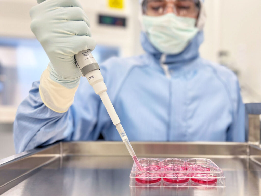
The cartilage biopsy sample is placed in a special nutrient-rich medium and secured in a temperature-controlled transport carrier. The carrier is transferred to an accredited clean-room laboratory facility, where the preparation process takes approximately 4 weeks.
Specialised, natural cartilage-forming cells are isolated and multiplied into millions of cells through an advanced biotechnological and strict quality-controlled process, usually over 3–4 weeks. After passing quality control tests, RECARTA® is released for implantation.
0
0
Second procedure – cartilage transplantation
0
1. Preparation
- The patient is prepared for arthroscopy, a minimally invasive procedure.
- The surgical site is cleaned, and local or general anaesthesia is administered.
2. Procedure (45 minutes)
- Small incisions, usually two or three, are made in the front part of the knee joint.
- The damaged cartilage area is prepared to receive the transplant. This involves removing damaged tissue and shaping the defect to fit the graft.
- The graft is then sized and trimmed to fit the defect precisely.
- Cartilage-forming cells are combined with a biological scaffold for improved attachment and proliferation.
- The tissue-engineered product is introduced back into the defect, and cartilage regeneration begins.
The joint is checked for any issues, such as graft movement or instability.

3. End of procedure and recovery
0
- The incision is closed with stitches.
- A dressing is applied, and the patient is moved to the recovery room.
- The knee is typically immobilised using a textile brace to protect the graft.
 PLASTIC SURGERY
 ORTHOPAEDIC SURGERY
 BARIATRIC SURGERY
 ABDOMINAL SURGERY
 UROLOGY SURGERY